![]()
Advanced wound care for complicated wounds
Each time we have used SertaSil at the clinic, we have after only a few treatments been able to achieve a completely clean wound bed without necrosis, infection or inflammation. And the wounds have healed rapidly and effectively every time without any need for additional treatment.
— Dr. Alf Peder Læbro, Vet & Partner.

Rat wound healing model
- SertaSil was compared to Gentaxane and un-treated Controls in a rat model of wound healing.
- SertaSil and Gentaxane were applied daily until achieving a clean wound, i.e. free from necrosis, pus and fibrinogenous thickenings..
- Rats receiving SertaSil reached the clean wound stage after 3 days compared to 7 and 10 days for Gentaxane and Control, respectively.
- Rats receiving SertaSil reached full wound closure after only 14 days compared to 18 and 23 days for Gentaxane and Control, respectively.
- At Day 13 the wound surface area was significantly smaller in the SertaSil group compared to the other groups and the wounds were almost completely closed.
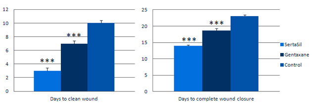
Effects of SertaSil in rat wound healing model. N= 15 per group. There were significant differences (P<0.001%) between all three groups.
SertaSil was compared to Gentaxane (gentamicin, L-tryptophan and zinc sulphate in a polymethylsiloxane powder) and Controls (untrated) in a rat model of wound healing. SertaSil and Gentaxane were applied every 24 hours until reaching a clean wound, i.e. free of necrosis, pus and fibrinogenous thickenings.
The SertaSil group reached the stage of a clean wound after 3 days compared to 7 and 10 days for Gentaxane and Control, respectively (see figure above). This effect resulted in a general acceleration of the wound healing process such that the SertaSil-group reached full wound closure after only 14 days compared to 18 and 23 days for Gentaxane and Control, respectively.
The wound surface area was measured at Day 13. It was 5.2±0.4 sq.mm in the SertaSil group compared to 38.0±0.4 and 95.7±2.9 sq. mm for Gentaxane and Control, respectively.
Wound smears were taken at regular time points and analysed for presence of immune cells. The table below shows the presence of neutrophils, monocytes and lymphocytes in the wound at 4 time points. The Gentaxane and the Control groups were essentially similar. In contrast, the SertaSil group had almost 30% lower levels of neutrophils whereas the levels of monocytes and lymphocytes were elevated by around 600% and 250%, respectively.
Neutrophils are the predominant cell type in wounds for the first 48 hours after injury and are known to protect the wounds from invading pathogens, cleanse the wound site of necrotic matter and release inflammatory mediators. In contrast, macrophages and lymphocytes appear to be important for the subsequent rebuilding and they have been shown to be essential to wound healing (DiPietro 1995; Mirza et al. 2009; Peterson et al. 1987). It is therefore of interest that the neurophils are lower in the SertaSil group, whereas both the macrophages and the lymphocytes are highly elevated compared to the Gentaxane and the Control groups. These data therefore support that SertaSil (MPPT) aids the immune system in gaining control of the wound such that it more rapidly can proeed to the following wound healing phases.
| Leucocytes in wound at different time points | SertaSil n=15 (mean ± SEM) | Gentaxane n=15 (mean ± SEM) | Control n=15 (mean ± SEM) |
| Neutrophils | |||
| 6h | 70.0 ± 0.39 | 95.0 ± 0.26 | 98.0 ± 0.20 |
| 12h | 75.0 ± 0.24 | 94.0 ± 0.17 | |
| 24h | 65.0 ± 0.35 | 94.0 ± 0.39 | 96.0 ± 0.22 |
| 48h | 60.0 ± 0.34 | 92.0 ± 0.24 | 88.0 ± 0.41 |
| Monocytes | |||
| 6h | 5.0 ± 0.20 | 1.0 ± 0.0 | 1.0 ± 0.01 |
| 12h | 5.0 ± 0.28 | 2.0 ± 0.17 | |
| 24h | 10.0 ± 0.44 | 2.0 ± 0.26 | 1.0 ± 0.14 |
| 48h | 12.0 ± 0.29 | 1.0 ± 0.14 | 2.0 ± 0.35 |
| Lymphocytes | |||
| 6h | 25.0 ± 0.35 | 4.0 ± 0.26 | 1.0 ± 0.17 |
| 12h | 20.0 ± 0.26 | 4.0 ± 0.26 | |
| 24h | 20.0 ± 0.28 | 3.0 ± 0.22 | 2.0 ± 0.17 |
| 48h | 21.0 ± 0.28 | 6.0 ± 0.26 | 8.0 ± 0.20 |
Presence of neutrophils, monocytes and lymphocytes in the wound at 4 time points.
![]()
References
- Dovi, J.V., He, L. and DiPietro, L.A. (2003) Accelerated wound closure in neutrophil-depleted mice. Journal of Leukocyte Biology 73:448-455.
- Mirza R, DiPietro LA, Koh TJ. (2009) Selective and specific macrophage ablation is detrimental to wound healing in mice. Am J Pathol. 2009 175(6):2454-62.
- Peterson JM, Barbul A, Breslin RJ, Wasserkrug HL, Efron G. (1987) Significance of T-lymphocytes in wound healing. Surgery 102(2):300-5.